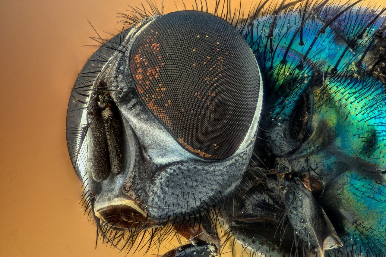
What do a housefly, a squid, and a human being have in common? All three see the world through eyes that use lenses. That shared trick, bending light through a curved, transparent structure, shows up across the animal kingdom in wildly different forms. Some lenses are tiny and fixed. Others flex and reshape dozens of times a minute. A few animals skip lenses entirely. This article looks at how lens-based eyes actually work, where they came from, how they fail, and what humans have built to mimic them.
What Counts as an Eye With a Lens?
A lens is a transparent structure that bends light so it converges into a focused image. In a camera-type eye, a single lens projects that image onto a sheet of photoreceptors at the back. In a compound eye, hundreds or thousands of tiny lens units each capture a small slice of the scene. (Google lens how to search using a picture)
Not every image-forming eye uses a lens, and that distinction matters. The nautilus, a shelled cephalopod, sees through a simple pinhole with no lens at all. Some scallops and certain deep-sea fish use curved mirrors instead. These are genuine eyes, just built on different optical principles. So “eyes with lenses” describes a category, not a universal rule.
The Vertebrate Lens: One Structure, Many Adjustments

The vertebrate eye focuses with a single lens, and that lens is a strange piece of biological engineering. It is made mostly of crystallin proteins packed into tightly ordered fibre cells. It contains no blood vessels. It gets its nutrients from the fluids around it, the aqueous humour in front and the vitreous humour behind. Because it has no blood supply, it also has no immune patrol, which is part of why the eye maintains special immune privileges.
How Accommodation Works
Focusing happens in two main ways across vertebrates:
| Animal group | Focusing method | What changes |
|---|---|---|
| Mammals and birds | Accommodation by reshaping | Ciliary muscles alter lens curvature |
| Fish and amphibians | Accommodation by moving | The lens shifts position rather than changing shape |
| Insects (compound eyes) | Fixed facet optics | Each ommatidium samples a fixed direction |
In practice, this means a human reading a book relies on muscles squeezing a flexible lens into a rounder shape. A fish instead slides its rigid lens back and forth. Same goal, different machinery.
Why Transparency Is So Fragile
Lens clarity depends on proteins staying in precise order. When that order breaks down, proteins clump and the lens clouds over. That clouding is cataract, and it remains one of the leading causes of blindness worldwide. The biology is unforgiving: a lens that loses its architecture cannot simply rebuild itself, because the oldest fibre cells at the centre are never replaced.
Compound Eyes: Thousands of Lenses Working Together
An insect’s compound eye is built from repeated units called ommatidia. Each ommatidium carries its own cornea and a crystalline cone that acts as a lens, funnelling light onto a small cluster of photoreceptor cells. A dragonfly may pack tens of thousands of these units into one eye.
Two optical arrangements matter here:
- Apposition eyes: each ommatidium forms its own isolated image point. This suits bright daylight and gives sharp resolution.
- Superposition eyes: a clear gap between lens layers lets light from many facets combine onto a single receptor. This is common in nocturnal insects and crustaceans and trades sharpness for sensitivity.
That trade-off is the same one photographers make when they open a lens aperture wide in dim light. Compound eyes solve it with architecture rather than mechanics.
Two Separate Roads to the Camera Eye
One of the most striking facts about lens eyes is that they evolved more than once. Camera-type eyes with lenses appear to have arisen independently in vertebrates and in cephalopods such as squid and octopus. The end result looks similar from the outside: a single lens, an iris, a retina.
The wiring underneath is not the same. Vertebrate retinas are inverted, with nerve fibres and blood vessels sitting in front of the photoreceptors, so light passes through several layers before it is detected. Cephalopod retinas are arranged the other way, with photoreceptors facing the incoming light directly.
This difference fuels a popular argument that the octopus eye is “better designed” than the human eye. It is a fair talking point, but the functional cost of the inverted retina is genuinely debated among vision researchers. Treat the claim as an argument, not settled science.
How Crystallins Got Recruited From Other Jobs
Lens proteins did not appear out of nowhere. Many crystallins are closely related to proteins that do other work elsewhere in the body. Some resemble small heat-shock proteins, which help cells cope with stress. Others resemble metabolic enzymes, including lactate dehydrogenase and argininosuccinate lyase.
This idea is known as gene sharing, associated with the work of Joram Piatigorsky and Graeme Wistow from the 1980s onward. A gene that already made a stable, soluble protein could be borrowed for optical duty in the lens without losing its original function. In practical terms, evolution reused existing parts rather than inventing a new protein family from scratch. It is one of the clearest examples of biology being a tinkerer rather than an engineer.
What Lens Eyes Do Well, and Where They Struggle
Lens eyes deliver something pinhole and mirror eyes generally cannot: a bright, sharply focused image with adjustable depth of field. That combination supports predation, navigation, and fine colour discrimination.
The costs are real too:
- Bulk and weight. A lens takes space and adds mass to the head.
- Vulnerability. A lens can cloud, scratch, or detach.
- Maintenance. Clear proteins must stay clear for decades in long-lived animals.
Anyone who has handled optical equipment knows the maintenance problem firsthand. A smudged lens scatters light and softens contrast, and the same principle applies to a biological lens. If you wear glasses or handle camera optics, the practical rules for keeping a lens clean without wrecking its coating are worth knowing, and the same care habits transfer to any precision optical surface. (Clean projector lens safely without damaging coating)
When the Lens Goes Wrong: Cataract and Correction
Because lens transparency depends on ordered protein packing, even small disruptions can have outsized effects. Cataract develops when crystallin proteins aggregate and scatter light instead of transmitting it. Age is the biggest risk factor, but ultraviolet exposure, diabetes, smoking, and trauma all contribute.
The standard treatment is surgical removal of the clouded lens and replacement with an intraocular lens, or IOL. These are engineered optical lenses, sometimes with adjustable focusing power. Corrective spectacle lenses and contact lenses address a different problem, refractive error, by placing an artificial lens in front of the eye rather than inside it.
| Lens type | Where it sits | Main job |
|---|---|---|
| Biological crystalline lens | Inside the eye | Focuses light, adjusts for distance |
| Spectacle lens | In front of the eye | Corrects refractive error |
| Contact lens | On the cornea | Corrects refractive error, thinner optical path |
| Intraocular lens | Replaces the natural lens | Restores focus after cataract surgery |
Human-Made Lenses and the Eyes That Use Them
Engineered lenses have quietly reshaped how millions of people see. But they introduce their own quirks. Progressive spectacle lenses create distortion at the edges of the field. Contact lenses reduce peripheral vision slightly and demand careful hygiene. IOLs are fixed in most cases, so the eye loses its natural ability to fine-tune focus.
There is also a habit worth flagging: people often assume a blurry view means a dirty lens. Sometimes it does. Sometimes the problem is a scratched coating, a misaligned frame, or simple fatigue. Checking the simplest cause first saves a lot of unnecessary cleaning, and unnecessary cleaning is how coatings get damaged in the first place.
Looking at Vision From the Other Direction
Lens eyes are a biology topic, and the same optical logic powers the cameras and sensors people use daily, including the lens in a smartphone that identifies objects, text, and scenes. Understanding how a lens forms an image makes it easier to reason about what those systems can and cannot do. Image recognition tools process what a camera captures with the same principle an ommatidium uses: gather light, focus it, and interpret the pattern.
Conclusion
Eyes with lenses are one of biology’s most reused inventions, and also one of its most varied. Vertebrates reshape or slide a single protein-dense lens. Insects tile thousands of tiny lens units into a mosaic. Cephalopods arrived at a camera eye through an entirely separate evolutionary route, with retina wiring that runs the opposite way. The common thread is a simple physical trick: bend light so that scattered rays converge into a usable image. That trick is powerful enough to have evolved repeatedly, fragile enough to cloud with age, and elegant enough that humans copied it into spectacles, contact lenses, and surgical implants. Whether the lens belongs to a squid, a housefly, or a person recovering from cataract surgery, the principle holds steady.
Frequently Asked Questions
What is the difference between a lens eye and a compound eye?
A lens eye uses one curved transparent structure to focus an image onto a retina. A compound eye uses many separate lens units called ommatidia, each sampling a small part of the scene. Compound eyes tend to excel at detecting motion and wide fields of view, while single-lens eyes generally deliver sharper overall resolution.
Do all animals with eyes have lenses?
No. The nautilus uses a pinhole eye with no lens, and some scallops and deep-sea fish use mirror-based optics. These animals still form images, just through different physical mechanisms. Lens-based eyes are widespread but not universal.
Why does the lens become cloudy with age?
The lens is built from tightly ordered crystallin proteins and cannot replace its oldest cells. Over time, proteins can clump and scatter light instead of transmitting it, producing cataract. Ultraviolet exposure, diabetes, smoking, and eye injury raise the risk.
Did the octopus eye really evolve separately from the human eye?
Yes, the evidence strongly supports independent evolution of camera-type eyes in vertebrates and cephalopods. Their retinas are wired differently, with vertebrate retinas inverted relative to the incoming light. The claim that one design is objectively superior remains debated rather than settled.
What does gene sharing mean in the lens?
Gene sharing describes how a gene already used for one job gets recruited for a second one. Many crystallins are closely related to stress proteins or metabolic enzymes, and they took on a structural, light-transmitting role in the lens while keeping their original function elsewhere.
Can a damaged lens coating be repaired?
Usually not. Anti-reflective and hydrophobic coatings are thin films, and once they wear through, the damage is permanent. Prevention works better: rinse loose grit away first, use a clean microfibre cloth, avoid dry wiping, and skip harsh solvents. The same gentle approach protects camera optics and eyeglass lenses alike.
That convergence is why the lens remains such a productive subject for study. Researchers can compare crystallin proteins across species to trace how the same molecular building blocks were repurposed again and again, and optical engineers can borrow those lessons when designing lightweight, durable lenses for everything from surgical microscopes to space telescopes. The eye is not a finished design but a snapshot of evolution mid-tinker, and every lens we build is a small echo of that long experiment.