A dog runs across the yard, yelps once, and suddenly won’t put weight on a back leg. It looks like a classic sports injury, the kind an athlete gets from a hard cut or a bad landing. But when the vet gives the diagnosis, many owners hear something unexpected: this was probably building for months.
That gap between what a canine ACL rupture looks like and what it actually is trips up a lot of dog owners. The injury that appears out of nowhere is usually the final stage of a slow, quiet degeneration inside the knee joint. Understanding that difference changes how you read the early warning signs, how you weigh treatment options, and what you can realistically expect afterward.
First, a Naming Correction That Matters
In people, the ACL is the anterior cruciate ligament, one of the two crossing ligaments that hold the knee steady. In dogs, the same structure is properly called the cranial cruciate ligament, or CCL. Veterinary professionals use CCL almost exclusively, while owners and general web searches stick with ACL. The two terms describe the same anatomy, so don’t let the vocabulary confuse you when talking to your vet.
What matters more is the anatomy itself. The cranial cruciate ligament sits inside the stifle joint, which is the dog’s equivalent of a human knee. It connects the femur to the tibia and stops the tibia from sliding forward when the leg bears weight. It also limits internal rotation of the lower leg. When that ligament fails, the joint loses its brake, and every step becomes a small instability event that grinds down cartilage.
Why Dog Ruptures Are Usually Not “Accidents”
Human ACL tears are typically acute, non-contact athletic injuries. A soccer player plants a foot, the knee buckles, and the ligament snaps in an otherwise healthy joint. Dogs follow a different script.
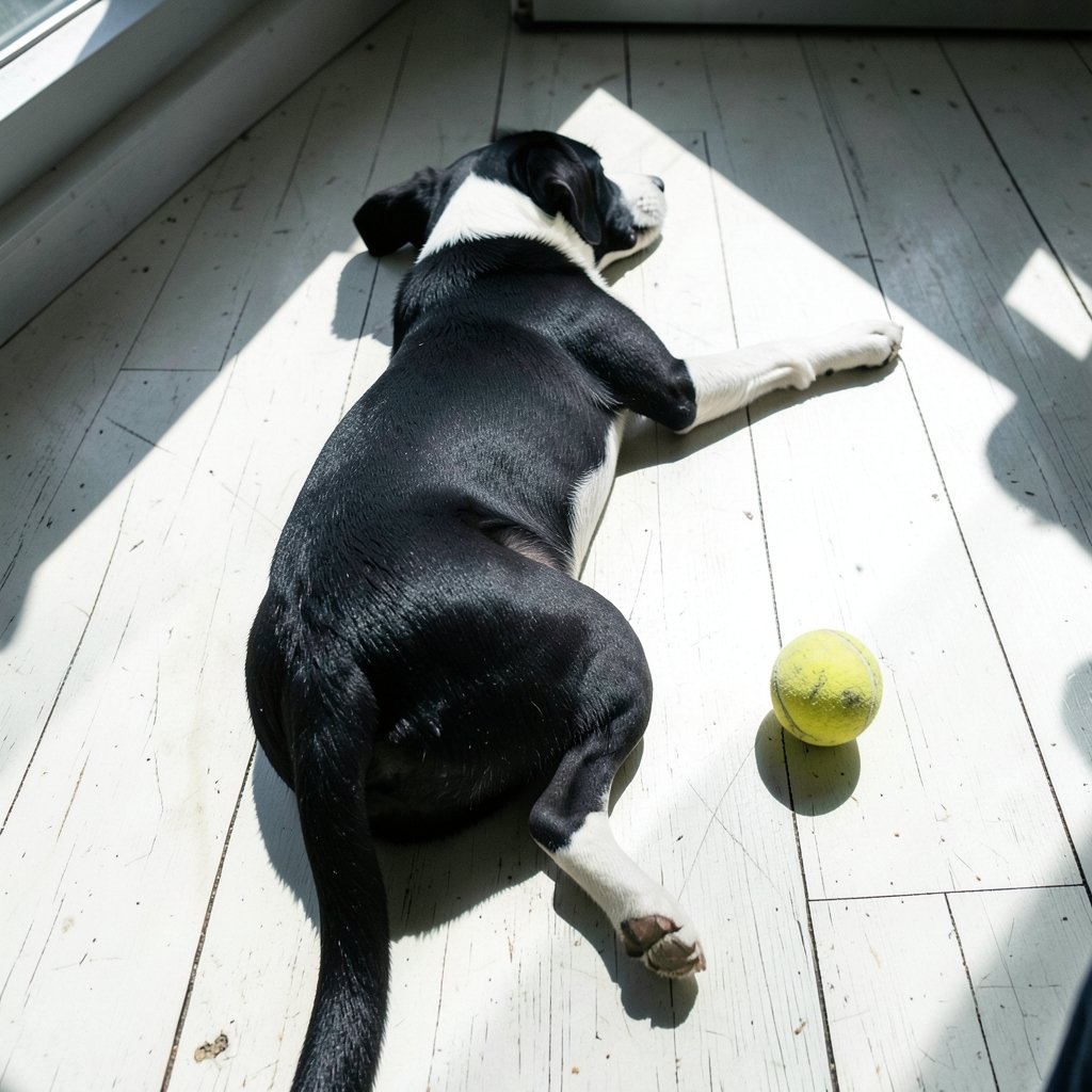
In dogs, the typical pattern is degenerative and progressive. The ligament slowly weakens, develops partial tears, and eventually ruptures completely. Often there is only minor trauma involved, or none at all. A dog can be walking down a hallway and go suddenly lame on a ligament that was already badly compromised. This is why veterinarians refer to the whole process as cranial cruciate ligament disease rather than a simple tear.
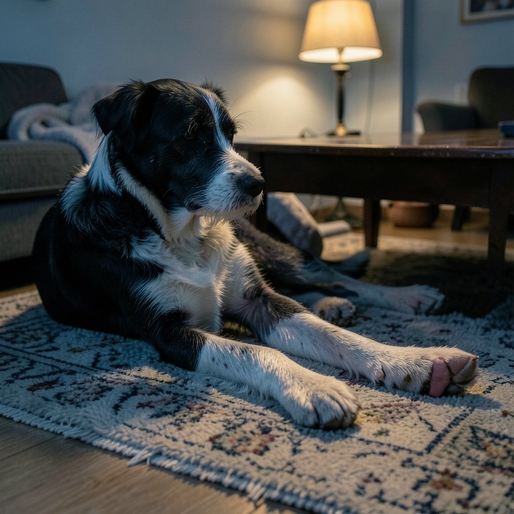
That distinction has real consequences. Owners who wait for a dramatic incident before taking limping seriously may miss months of partial tearing, during which arthritis quietly sets in. A dog that occasionally “skips” a step on a hind leg, or that sits with the affected leg tucked out to the side, is showing a pattern worth investigating.
Who Is at Risk?
Cranial cruciate ligament disease is one of the most common causes of hind-limb lameness in dogs, particularly in medium and large breeds. Several factors are consistently associated with higher risk:
| Risk Factor | What the Evidence Suggests |
|---|---|
| Increasing age | Risk rises as ligaments accumulate wear |
| Higher body weight and obesity | Extra load accelerates ligament stress and arthritis |
| Neutering, especially early | Associated with higher risk in several studies |
| Breed predisposition | Labrador Retrievers, Rottweilers, and Newfoundlands appear more often |
Effect sizes vary between studies, so treat any specific percentage you see online with caution. The pattern is reliable; the exact numbers are not. For owners, the practical takeaway is that body condition is the factor you can actually control. Keeping a predisposed dog lean is one of the few levers that clearly helps.
Recognizing the Signs Before a Full Tear
The classic presentation is a dog that suddenly holds up a hind leg after exercise. But the more useful skill is spotting the partial-tear phase. Watch for these patterns:
- Intermittent lameness that appears after activity and then seems to resolve.
- A shifted sitting position, with the affected leg extended outward rather than tucked under the body.
- Reluctance to rise from lying down, especially after rest.
- Muscle wasting on the affected thigh, which develops surprisingly fast.
- A clicking sound when the dog walks, sometimes audible across a room, which may indicate meniscal involvement.
That last point deserves attention. Medial meniscal tears commonly accompany cruciate rupture, and they are a frequent reason a dog stays lame even after surgery. If your dog had surgery and is still limping weeks later, meniscal injury is one of the first things a surgeon will consider.
How Vets Confirm the Diagnosis
Diagnosis is built on two pillars: the physical exam and imaging. On the exam table, veterinarians look for two classic signs. The cranial drawer test checks whether the tibia can be pushed forward relative to the femur. The tibial compression test looks for the same abnormal motion while the leg is flexed. Both indicate instability, though muscle tension and patient cooperation can affect results, which is why sedation is sometimes used.
Radiographs serve a supporting role. They don’t show the ligament itself, but they reveal joint effusion and the secondary osteoarthritis that develops with chronic instability. Advanced imaging like MRI or CT exists but is used less often in routine cases because the physical exam usually settles the question.
Treatment: What the Options Actually Involve
Surgical stabilization is generally recommended for medium and large dogs. The logic is straightforward: the ligament won’t heal on its own, and ongoing instability drives arthritis forward. Several techniques are in common use.
Tibial plateau leveling osteotomy (TPLO) reshapes the top of the tibia so the joint no longer relies on the ligament to resist forward sliding. Tibial tuberosity advancement (TTA) achieves a similar goal through a different geometric approach. Lateral fabellar suture and tightrope procedures use synthetic material to mimic the ligament’s restraint.
Here’s the honest part: comparative outcome data between these techniques are mixed, and study designs vary widely. Implying one method is universally superior overstates what the evidence shows. Technique choice often comes down to the surgeon’s training and experience, the dog’s conformation, and cost. Ask your surgeon why they favor a particular approach for your dog specifically.
Conservative management, meaning rest, weight control, and physiotherapy, is mainly an option for small dogs or for dogs that aren’t good surgical candidates. For a large, active dog, nonsurgical care usually means accepting progressive arthritis and ongoing lameness.
The Arthritis Conversation Nobody Wants
Regardless of which treatment path you choose, progressive osteoarthritis in the affected stifle is common. This isn’t a failure of surgery; it’s the nature of a joint that has experienced instability. What you can influence is how fast it progresses.
Weight management is the key modifiable factor. Every extra pound on a dog with a compromised knee is load the joint has to absorb thousands of times a day. Physiotherapy, controlled exercise, and joint-supportive strategies all have a role, but none of them outrun excess body weight.
Owners often ask whether the other leg is at risk. It is. Dogs with cruciate disease on one side frequently develop it on the other, partly because they shift weight onto the healthy leg while compensating. That’s another reason early diagnosis and prompt treatment matter.
Practical Tips and Common Mistakes
A few things come up again and again in real cases, and knowing them in advance saves frustration.
Mistake one: waiting to see if it gets better. Partial tears don’t resolve with time; they progress. A week of rest that produces improvement can create false confidence.
Mistake two: skipping the rehab plan. Surgery repairs mechanics, not muscle. Dogs that follow a structured physiotherapy program typically return to function more smoothly than those that go straight back to the couch and then straight back to the dog park.
Mistake three: returning to full activity too fast. The bone cuts in procedures like TPLO need time to heal. Letting a dog bolt after a squirrel at week four is a common cause of setbacks.
What actually works: strict rest during recovery windows, leash-only walks for the full prescribed period, non-slip flooring at home, weight control started before surgery rather than after, and a written rehab schedule you actually follow. Slippery hardwood is a genuine hazard for a three-legged dog learning to walk again.
Conclusion
Canine ACL rupture is one of those diagnoses that reframes how you think about your dog’s movement. The dramatic yelp gets the attention, but the real story is usually a ligament that has been failing quietly for months. That’s good news in a way, because it means attentive owners can catch the problem during the partial-tear phase, when intervention tends to go better. Keep your dog lean, take intermittent hind-leg lameness seriously, and ask direct questions about surgical options and rehab timelines. For a deeper look at canine orthopedic care and recovery planning, visit OCCCY. Your dog’s knee will thank you for the attention long before anything snaps.
What Is the Difference Between a Dog’s ACL and CCL?
They are the same structure. The cranial cruciate ligament (CCL) is the anatomically correct veterinary term for the ligament in a dog’s stifle joint. ACL is the human term for the equivalent ligament in people. Veterinarians use CCL because it describes the ligament’s position relative to the front of the joint, and because the disease process in dogs differs meaningfully from the acute athletic injuries seen in humans.
Can a Dog’s Torn ACL Heal on Its Own?
No. The cranial cruciate ligament does not have a meaningful capacity to heal once ruptured, largely because it sits inside a joint with limited blood supply to the tissue. Small dogs can sometimes be managed without surgery through rest, weight control, and physiotherapy, but the joint remains unstable and arthritis progresses. For medium and large dogs, surgical stabilization is generally recommended.
How Long Is Recovery After Cruciate Surgery?
Recovery timelines vary by procedure and by dog, but most surgeons prescribe roughly eight to twelve weeks of restricted activity, with gradual increases in leash walking. Bone-healing procedures like TPLO require time for the surgical site to consolidate. Physiotherapy typically runs alongside this period. Full return to unrestricted activity, including off-leash running, usually comes later than the initial restriction window ends.
Why Is My Dog Still Limping After Surgery?
Persistent lameness after cruciate surgery has several possible causes. Medial meniscal tears are among the most common and frequently require a second procedure. Other possibilities include incomplete healing, infection, implant complications, or simply insufficient rehab and muscle rebuilding. Any dog still lame weeks after surgery should be reassessed rather than kept on rest indefinitely.
Is My Other Dog Leg Going to Rupture Too?
It’s a real concern. Dogs that develop cruciate disease on one side have a meaningfully elevated risk on the other, partly because they load the healthy leg more heavily during compensation. Keeping body weight controlled, maintaining muscle strength through physiotherapy, and addressing lameness early are the most practical ways to reduce that risk. Owners should watch the opposite leg closely in the months after treatment.
What Can I Do to Lower My Dog’s Risk?
Focus on what’s modifiable. Keep your dog at a lean body condition, since obesity is one of the strongest controllable risk factors. Maintain steady, appropriate exercise rather than sporadic intense bursts. Consider non-slip flooring for older or predisposed dogs. If you’re choosing a breed known for cruciate issues, go in aware. Age, neutering status, and breed are fixed factors, but weight and conditioning are not.
A final note on timing. The single most useful thing an owner can do is act on the first episode of intermittent lameness rather than the first full tear. A dog examined during the partial-tear phase has more options, a shorter recovery, and a better long-term joint than one examined after months of compensation. When in doubt, book the appointment. An exam costs far less than a delayed diagnosis.