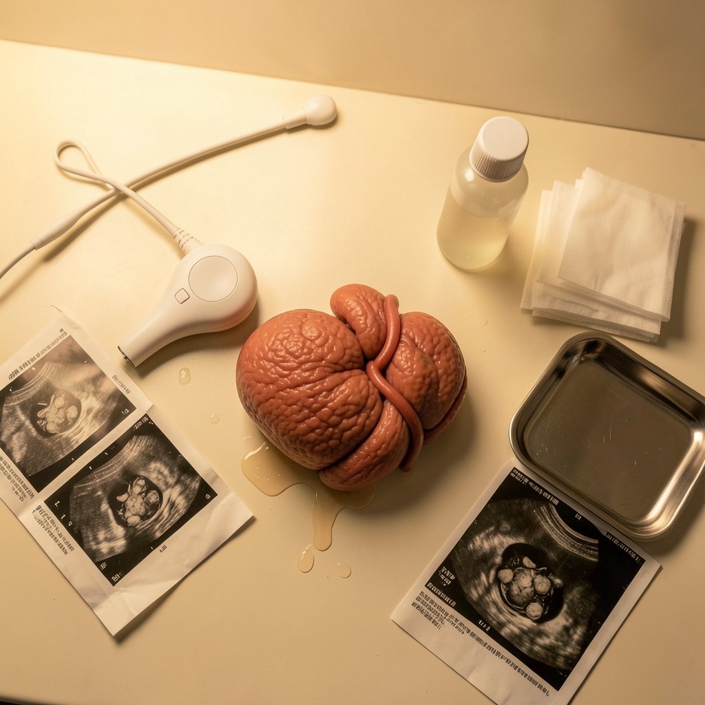
What if the lifeline between a baby and the placenta took a wrong turn before it ever reached home? That is essentially what happens in velamentous cord insertion (VCI), a placental anomaly where the umbilical cord attaches to the fetal membranes instead of the fleshy center of the placenta. The vessels then travel unprotected through the membrane before reaching the placental disk. It sounds alarming, and in some cases it genuinely is. But most pregnancies with this finding still end with a healthy baby. The key is knowing what to watch for, how it is diagnosed, and which risks actually matter.
What Is Velamentous Cord Insertion?
In a typical pregnancy, the umbilical cord inserts directly into the placental disk, where the vessels are cushioned by a thick layer of tissue. In VCI, the cord inserts into the chorioamniotic membranes instead. From there, the umbilical vessels run through the membranes without the protective coating of Wharton’s jelly, the gelatinous substance that normally insulates and shields the cord.
Those exposed vessels are the heart of the problem. Because they lack that cushioning, they are more vulnerable to compression and rupture. This distinction matters more than most expectant parents realize, and it shapes how clinicians monitor the pregnancy from the second trimester onward.
How Common Is It, Really?

Prevalence figures vary widely depending on how a study counted cases. Broadly, VCI is reported in roughly 1% of singleton pregnancies and about 1.5% of twin pregnancies, with higher rates in monochorionic twins, who share a single placenta. These numbers are approximate because some studies diagnose at ultrasound while others rely on findings at delivery, and antenatal detection rates are often low. That means registry-based figures may undercount the condition.
The practical takeaway: VCI is uncommon but not rare. If it shows up on a scan, it is a known finding that obstetric teams encounter regularly.
Why the Cord Insertion Site Matters
The umbilical cord carries oxygen and nutrients to the fetus and removes waste. When the insertion point is off-center, the vessels take a longer, more exposed route. Three consequences follow:
- Vessel vulnerability. Unprotected vessels can be compressed during contractions or ruptured when membranes break.
- Association with vasa previa. VCI is the main risk factor for vasa previa, a condition where unprotected fetal vessels cross the internal cervical os. If those vessels tear during membrane rupture, the fetus can lose blood rapidly, which is a genuine emergency.
- Growth concerns. Some studies link VCI to fetal growth restriction and low birth weight, though findings are inconsistent across cohorts and the effect size is debated.
Risk Factors Linked to VCI
Observational studies have reported associations with several factors. These are associations, not proven causes:
| Risk Factor | Reported Findings |
|---|---|
| Multiple gestation (twins) | Higher rates, especially in monochorionic twins |
| Low-lying placenta or placenta previa | Repeatedly reported association |
| Advanced maternal age | Reported in several cohorts |
| In vitro fertilization (IVF) | Associated in observational data |
| Smoking | Reported association |
One important nuance: because placenta previa often coexists with VCI, researchers debate whether growth restriction is caused by the cord insertion itself or by the placental position. That uncertainty is worth holding onto when reading scary statistics online.
How Is VCI Diagnosed?
Prenatal diagnosis relies on obstetric ultrasound, typically with color Doppler to trace where the cord meets the placenta. A skilled sonographer can follow the cord and identify an insertion into the membranes rather than the disk.
In practice, though, VCI is frequently missed before delivery. Detection depends on the sonographer’s technique, the placenta’s position, and whether the condition is specifically looked for. Many cases are discovered only at birth when the delivering clinician inspects the placenta and cord.
This is where practical experience matters. If a pregnant person has a known risk factor, such as twins or a low-lying placenta, it is reasonable to ask whether the cord insertion was visualized during the anatomy scan. A simple question can prompt a closer look. For anyone documenting scans or appointments, tools like this guide on how to screen record iPhone steps audio tips fixes can help keep a clear record of what was discussed, though clinical decisions always belong with the care team.
What Happens After a Diagnosis?
There is no treatment for VCI itself. Management focuses on surveillance and planning for delivery. Guidelines generally recommend closer monitoring of fetal growth, often with serial growth scans, and careful planning if vasa previa coexists.
When vasa previa is present, clinicians commonly consider cesarean delivery before labor to avoid rupturing the membranes and the vessels crossing the cervix. For isolated VCI without vasa previa, many pregnancies are managed with routine or slightly enhanced monitoring, and vaginal delivery is often possible.
The single most important variable is whether vasa previa is also present. That combination is where the serious risk concentrates.
What the Outcomes Usually Look Like
Here is the reassuring part: most singleton pregnancies with isolated VCI have normal outcomes. The elevated risk is concentrated in cases with vasa previa, growth restriction, or prematurity. In other words, the diagnosis alone is not a verdict. It is a flag that prompts closer attention.
That said, the growth-restriction link deserves honest treatment. Findings across studies are inconsistent, and some of the risk may be driven by coexisting conditions like placenta previa. Expectant parents should ask their clinician what the monitoring plan means for their specific case rather than extrapolating from general statistics.
Common Mistakes and Practical Tips
A few patterns show up again and again in how people handle this diagnosis:
- Assuming the worst from a single statistic. Prevalence and outcome figures come from different study designs. A number pulled from a high-risk twin cohort does not describe a low-risk singleton pregnancy.
- Not asking whether the cord insertion was visualized. Many anatomy scans do not explicitly document it. Asking directly is reasonable and low-effort.
- Skipping growth scans because the diagnosis “seems minor.” Serial growth monitoring is the main tool for catching fetal growth restriction early.
- Confusing VCI with vasa previa. They are related but distinct. VCI describes where the cord attaches; vasa previa describes vessels crossing the cervix. The second is the more dangerous finding.
- Relying on forums over the care team. Online communities can offer comfort, but management decisions depend on individual placental position, fetal growth, and gestational age.
For anyone who wants to build a well-organized record of appointments, scan dates, and questions, a systematic approach helps. The same discipline that goes into choosing a microphone that fits your actual recording needs applies here in spirit: match the tool to the job, and do not over-engineer. Keeping a simple log of scan findings and growth percentiles is usually enough.
Questions Worth Asking Your Care Team
Specific questions tend to produce specific answers. Consider asking:
- Was the cord insertion site visualized during my anatomy scan?
- Is there any evidence of vasa previa, and if so, how is it being monitored?
- What is the plan for growth surveillance, and how often will scans happen?
- Does the placental position change the delivery plan?
- At what point would cesarean delivery be recommended?
These questions move the conversation from general statistics to the particulars of one pregnancy, which is where decisions actually get made.
When VCI Coexists With Other Conditions
The clinical picture shifts when VCI appears alongside other findings. Twin pregnancies, particularly monochorionic twins, carry higher baseline rates and more complex monitoring. Placenta previa adds its own considerations around delivery timing and bleeding risk. In these combined scenarios, the care team is balancing several variables at once, and delivery planning often involves a maternal-fetal medicine specialist.
If a household is juggling appointments, work, and childcare, keeping logistics straight can feel like its own project. Some families use screen recording to capture telehealth visits or instructions, and guides like this one on screen record mac audio actually works explain how to get clean audio. It is a small practical step, but clear records reduce the chance of misremembering instructions.
The Bottom Line on Monitoring
VCI is a structural quirk of placental anatomy, not a disease with a cure. The clinical response is watchful: confirm the diagnosis, rule out vasa previa, track fetal growth, and plan delivery accordingly. Most isolated cases do well. The cases that need the most attention are the ones where exposed vessels sit over the cervix or where growth falls behind.
Conclusion
Velamentous cord insertion sounds frightening, and the exposed vessels it creates are a real reason for careful monitoring. But the diagnosis is not destiny. Roughly 1% of singleton pregnancies and about 1.5% of twin pregnancies involve VCI, and most isolated cases end with a healthy baby. The risk concentrates in pregnancies where vasa previa, growth restriction, or prematurity also appear. The most useful thing an expectant parent can do is ask whether the cord insertion was visualized, understand whether vasa previa is present, and keep up with growth scans. Clear communication with the care team turns a confusing finding into a manageable plan.
Frequently Asked Questions
Can velamentous cord insertion resolve on its own?
No. The cord insertion site is fixed once the placenta forms, so VCI does not resolve during pregnancy. What can change is the clinical picture around it, such as fetal growth patterns and placental position. Management focuses on monitoring those variables rather than expecting the insertion to change.
Is velamentous cord insertion the same as vasa previa?
They are related but distinct. VCI describes where the umbilical cord attaches to the membranes instead of the placental disk. Vasa previa describes unprotected fetal vessels crossing the internal cervical os. VCI is the main risk factor for vasa previa, but most people with VCI do not have vasa previa.
Does VCI always mean a cesarean delivery?
No. Isolated VCI without vasa previa is often managed with enhanced monitoring, and vaginal delivery is frequently possible. Cesarean delivery is more commonly considered when vasa previa is present, because rupturing the membranes could tear the exposed vessels. The delivery plan depends on the full clinical picture.
Can VCI cause fetal growth restriction?
Some studies report an association between VCI and fetal growth restriction or low birth weight, but findings are inconsistent across cohorts. Researchers also debate whether the growth risk comes from the cord insertion itself or from coexisting conditions such as placenta previa. This is why serial growth scans matter more than any single statistic.
How is VCI detected during pregnancy?
Prenatal detection uses obstetric ultrasound, usually with color Doppler to trace where the cord meets the placenta. Detection rates vary, and many cases are found only at delivery when the placenta and cord are examined. Asking whether the insertion site was visualized during the anatomy scan is a reasonable step.
Does VCI affect the baby after birth?
For most babies with isolated VCI, outcomes are normal. Risks are concentrated in pregnancies complicated by vasa previa, growth restriction, or prematurity. Babies who grow well and are delivered at term generally do not face long-term effects from the cord insertion itself, though each case is assessed individually by the care team.
One final point deserves emphasis: the anxiety surrounding a VCI diagnosis often outpaces the actual risk. Reading a single study or forum thread can make a rare complication feel imminent, but the statistics are clear that most isolated cases proceed without incident. If monitoring reveals stable growth and no vasa previa, there is usually little reason to alter everyday life. Staying active, attending scheduled scans, and reporting any bleeding or unusual symptoms promptly are the practical steps that matter most. Everything else, from delivery timing to surveillance intervals, is a decision best made with the care team, informed by the specifics of one pregnancy rather than the averages of many.